SATO Wataru Laboratory
The atypical social brain network in autism: Advances in structural and functional MRI studies
(Sato*, & Uono* (* equal contributors): Curr Opin Neurol)
Purpose of review
To review advances in structural and functional magnetic resonance imaging (MRI) studies regarding the neural underpinnings of social atypicalities in autism spectrum disorder (ASD).
Recent findings
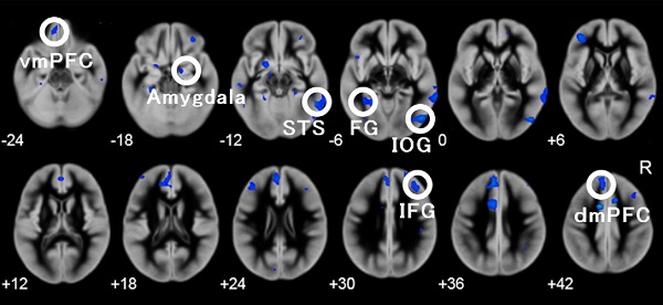
According to the hypothesis that the social brain network, which includes brain regions, such as the amygdala and superior temporal sulcus, may be atypical in ASD, recent structural MRI studies have identified regional gray matter volume abnormalities in the social brain regions in ASD groups compared with the typically developing (TD) groups.
Studies evaluating gray matter volume covariance and white matter volume/integrity suggested network-level abnormalities associated with the social brain regions.
Recent functional MRI studies assessing resting-state neural activity showed reduced functional connectivity among the social brain regions in individuals with ASD compared with TD groups.
Similarly, task-based functional MRI studies recently revealed a reduction in regional activity and intra-regional functional coupling in the social brain regions during the processing of social stimuli in individuals with ASD.
Summary
These structural and functional MRI studies provide supportive evidence for the hypothesis that an atypical social brain network underlies behavioral social problems in ASD.
Return to
Recent Research.
Return to
Main Menu.